Tooth Explorer 3D — Interactive Dental Anatomy Platform
Context
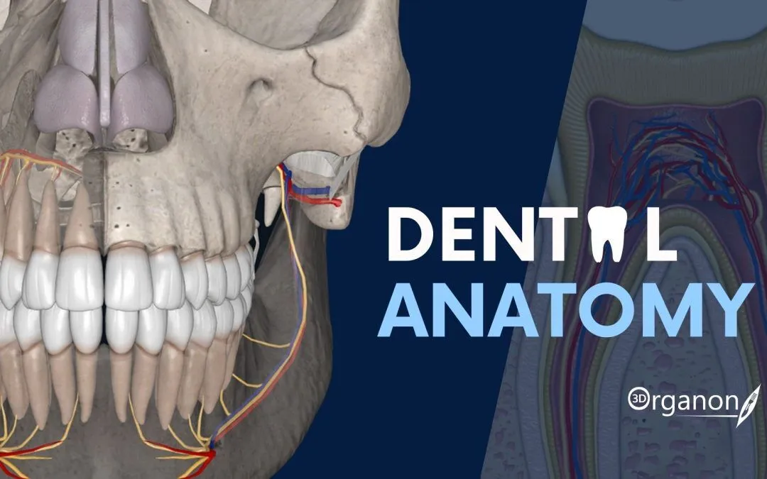
Tooth Explorer 3D is a specialized visualization tool created to support dental education and clinical orientation. Unlike static atlases, it provides a fully interactive 3D environment where dental students and professionals can examine teeth, occlusion, and surrounding structures in detail. Its role is primarily academic, but it also helps clinics in patient communication by demonstrating morphology and treatment concepts in an accessible format. The tool is not a diagnostic system; rather, it is an aid for understanding spatial relations in dental anatomy.
Technical Profile
| Area | Details |
| Platform | Windows desktop; VR-ready configuration optional |
| Dental focus | Tooth morphology, occlusion, surface details, anatomical orientation |
| Core modules | 3D tooth library, rotation/zoom tools, annotation and labeling |
| Interop | Standalone viewer; no direct PACS/EMR linkage |
| Imaging | Predefined 3D datasets; not based on clinical DICOM scans |
| Security | No patient data; purely educational content |
| Multisite | Deployed in classrooms, labs, or standalone PCs |
| Backup/DR | Not applicable; dataset is fixed |
| Licensing | Academic/free edition; commercial license adds extended models |
Scenarios (Dental-Specific)
– A preclinical course uses Tooth Explorer 3D to let first-year students explore molars and premolars before wax modeling.
– A university anatomy lab installs the tool on multiple PCs, providing consistent access to the same dataset.
– A clinic applies it in patient consultations, showing 3D occlusion views to explain orthodontic or restorative treatment plans.
Workflow (Admin View)
1. Install the software on a Windows workstation or classroom PCs.
2. (Optional) Connect VR headsets for immersive interaction.
3. Load the included anatomy dataset.
4. Configure annotation sets and labeling for teaching consistency.
5. Provide user orientation sessions for navigation and zoom functions.
6. Keep installation images updated across multiple lab stations for uniform access.
Strengths / Weak Points
Strengths
High-quality 3D tooth models that aid spatial learning.
Low barrier to entry — fixed dataset, no complex setup.
Useful for both academic instruction and patient-facing explanations.
Optional VR mode adds engagement for classrooms and workshops.
Weak Points
No integration with PACS or real clinical imaging.
Dataset is static; cannot load patient-specific cases.
Requires capable hardware for smooth rendering, especially in VR mode.
Limited assessment features compared to commercial teaching suites.
Why It Matters
Dental education often struggles with bridging the gap between textbook diagrams and real clinical images. Tooth Explorer 3D provides an intermediate layer, offering students and practitioners a chance to examine morphology and occlusion in an interactive way. For institutions without access to more expensive VR platforms, it represents a practical, lightweight entry point into 3D dental anatomy teaching. In clinics, its role as a visualization aid can improve patient understanding and acceptance of proposed treatments.

