Oral Histology Atlas (Free) — Digital Reference for Dental Tissues
Context
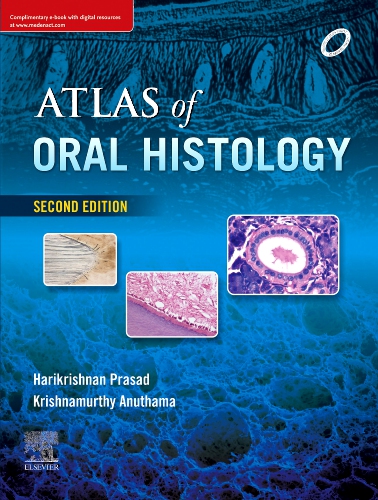
Oral Histology Atlas (Free) is a teaching resource that compiles high-resolution slides of oral tissues into a structured digital format. Instead of flipping through printed atlases or microscope slides, students and instructors can navigate a curated set of images that highlight enamel, dentin, pulp, cementum, and supporting structures. The tool was created mainly for dental faculties and training labs, where histology is a core subject but access to physical specimens is sometimes limited. While not a diagnostic system, it simplifies the way learners approach microscopic anatomy, making complex structures easier to identify and compare.
Technical Profile
| Area | Details |
| Platform | Windows and web-based editions, depending on distribution |
| Dental focus | Microscopic anatomy of oral and dental tissues |
| Core modules | Image library, labeling tools, search by tissue type, annotation mode |
| Interop | Standalone educational viewer, no link to EMR/PACS |
| Imaging | Preloaded histology slides (JPEG/PNG), not based on DICOM |
| Security | Educational data only, no patient records |
| Multisite | Suitable for classrooms, labs, and remote teaching platforms |
| Backup/DR | Simple filesystem copies; dataset is static |
| Licensing | Free edition; some distributions offer extended premium libraries |
Scenarios (Dental-Specific)
– A histology course uses the atlas to replace microscopes during introductory labs, letting students zoom into enamel and dentin structures on screen.
– Faculty upload the atlas to a classroom projector and annotate structures live during lectures.
– Distance-learning programs distribute the free edition so students can review tissues at home without needing lab access.
Workflow (Admin View)
1. Install the software or provide browser access, depending on version.
2. Load the default library of histology slides included with the free edition.
3. Organize content by course modules — enamel week, pulp week, etc.
4. Train students on navigation (zoom, labeling, search by tissue).
5. Share annotated slide sets with faculty notes for review sessions.
6. Keep software and slide packages consistent across all lab machines or virtual classrooms.
Strengths / Weak Points
Strengths
Free and easy to distribute to large student groups.
High-quality images that highlight essential dental tissue features.
Labeling and annotation tools useful for teaching and exams.
Reduces dependency on microscopes and fragile glass slides.
Weak Points
Static library — cannot expand with institution-specific images unless upgraded.
Limited to 2D images, no 3D histology or interactive models.
Not connected to clinical imaging or case studies.
Free versions may vary in slide count and resolution.
Why It Matters
Histology is one of the foundations of dentistry, but physical slide sets and microscopes are not always available in sufficient numbers. Oral Histology Atlas (Free) lowers this barrier, providing reliable, reusable images that make teaching and self-study easier. For institutions with limited budgets, it keeps learning consistent across classrooms and even supports remote education. While advanced research requires more comprehensive systems, this free atlas still plays a key role in making oral histology accessible, visual, and easier to understand.

